SmartRadReports is a set of web applications for structured reporting in nuclear medicine and radiology. You enter what you see using fixed options, a polar plot or a skeleton map, and the application writes the findings and the impression for you to copy into your reporting system.
In clinical use
The Nuclear Cardiology application has been in use for about three years and has been tested by multiple doctors.
How a report is made
- Open an application from your accountEach application opens from its page on this site. A timer in the application header shows how much of your session is left.
- Enter the findingsScore the polar plot, describe a nodule or click a bone on the skeleton map. Choices come from fixed lists, so the same finding is described the same way every time.
- Read the generated reportThe application writes the findings and the impression from your entries, using one consistent set of rules.
- Copy itOne button copies the whole report, ready to paste into your reporting or dictation system. To change the wording, edit it after you paste.
- Come back for the follow-upEvery report has a report ID. Use it to find the report again. In Thyroid Ultrasound and Bone Scan, the next exam starts with the earlier findings already in place.
The applications
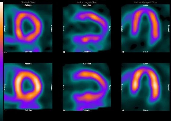
Nuclear Cardiology
Structured reports for gated myocardial perfusion studies, built around the 17-segment polar plot.
- Stress and rest polar plots, each segment scored from 0 (normal) to 4 (absence of uptake) by clicking
- Segments coloured by LAD, RCA and LCX territory
- Summed stress, rest and difference scores calculated for you
- LV size, TID, wall motion and thickening, and LVEF entered directly or calculated from EDV and ESV
- Coronary calcium score and myocardial blood flow by vessel, with flow reserve calculated
- Pull Old Report lists the reports from your institution so you can reopen and update one
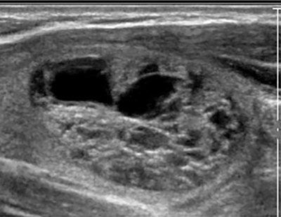
Thyroid Ultrasound
A structured description of the thyroid gland, nodules and lymph nodes, with risk assessment and recommendations.
- ATA or ACR TI-RADS, set for your institution; TI-RADS points and TR level calculated
- Up to four nodules in each lobe and the isthmus, and four lymph nodes per side
- Composition, echogenicity, margins, calcifications, shape and extrathyroidal extension from fixed lists
- Significant change in size worked out from the prior measurements
- A follow-up or FNA recommendation suggested for each nodule, which you can change
- Earlier exams of the same nodules shown when you log a new session
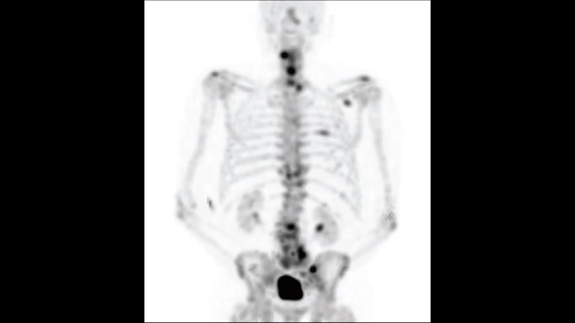
Bone Scan
Structured reports for whole-body bone scans, entered on an interactive skeleton map.
- Click bones on a zoomable anterior skeleton map to mark each lesion
- Assessments: metastasis, degenerative arthritis, healing fracture and osteomyelitis
- Certainty, interval change and a recommendation recorded for each lesion
- Follow-up exams start with the earlier lesions already marked, ready to update
- Every exam in a series shown side by side, lesion by lesion
Patient privacy
The applications do not ask for a patient name, medical record number, accession number or date of birth, and none of these is stored. What is saved is the findings you enter, your user name and institution, and the report ID. In Nuclear Cardiology and Thyroid Ultrasound, the list of earlier reports only shows reports from your own institution.
Help
Nuclear Cardiology and Thyroid Ultrasound include a guided tour of the reporting screen. Open it with Tutorial in the application header. For anything else, use the contact form.
Disclaimer
This tool does not replace medical training, clinical judgement, or personal understanding of the literature. The generated report must be re-checked and should not be used alone to guide patient care.